CT TAVI Planning

CT imaging in TAVI to advance patient care

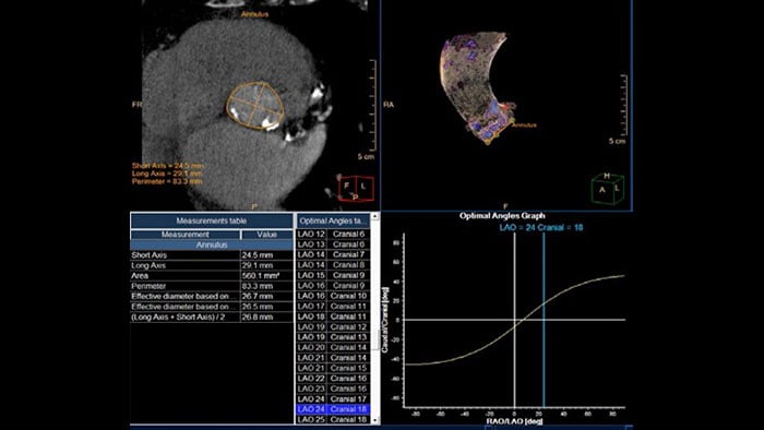
Provides 2D and 3D visualization and automated measurements designed to assist in proper TAVI-device sizing, on contrast-enhanced, prospectively ECG-gated axial or retrospectively gated helical CT images.

tavi planning thumbnail

Benefits

  • Area and diameter calculations for the aortic annulus, LVOT, sinotubular junction, sinus of valsalva and ascending aorta planes, as well as distance to coronary ostia from the annulus plane.
  • Tools such as inspection of minimal diameter, calcifications and vessel tortuosity, to assess the peripheral vessels along the access route.
  • 3D segmentation and visualization of aortic valve calcifications.

Our site can best be viewed with the latest version of Microsoft Edge, Google Chrome or Firefox.

Jūs pateksite į Lietuvos „Philips Healthcare“ svetainę.
Please select the checkbox

Svetainė skirta tik asmenims, naudojantiems medicinos priemones profesinėje veikloje, įskaitant sveikatos priežiūros specialistams, profesinę veiklą vykdantiems asmenims, atstovaujantiems sveikatos priežiūros įstaigą ar medicinos priemonėmis prekiaujančią organizaciją.

Ar esate asmuo, atitinkantis aukščiau nurodytus kriterijus?