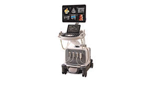
Abdominal Aortic Aneurysm Model
Advanced innovation in surveillance of abdominal aortic aneurysms
Image gallery
- Toggle view
Features

Increased diagnostic confidence
3D ultrasound with the AAA Model solution can be used to estimate the diameter and volume of an AAA with acceptable reproducibility and an improved agreement with CT, and with inter-operator reproducibility superior to that of a 2D ultrasound exam.

Improved patient experience
Philips AAA Model improves the patient experience by eliminating exposure to high levels of radiation and nephrotoxic contrast agents that CT Angiography (CTA) requires, while still providing clinicians with the necessary diagnostic information.

Lower cost of care
The low cost of ultrasound is one reason that it has become the preferred imaging modality for aneurysm screening and surveillance. The AAA model supports this important ultrasound imaging application.
The AAA model is compatible with:
-
EPIQ Elite
EPIQ Elite Elevate provides high-quality imaging and tailored clinical information to help clinicians deliver timely, confident answers to more patients worldwide. With advanced intelligence and an exceptional level of performance, EPIQ Elite meets the demands of today’s most challenging practices.
795098
Documentation
- Resources
-
Related stories
Footnotes
i Howard DP, Banerjee A, Fairhead JF, et al. Age-specific incidence, risk factors and outcome of acute abdominal aortic aneurysms in a defined population. British Journal of Surgery. 2015;102(8):907-915. doi:10.1002/bjs.9838. www.ncbi.nlm.nih.gov/pmc/articles/PMC4687424 Philips Abdominal Aortic Aneurysm Model is available in selected countries. Please consult your Philips representative for further details.
ii Bredahl K, et al. Three-dimensional ultrasound evaluation of small asymptomatic abdominal aortic aneurysms. European Journal of Vascular and Endovascular Surgery. 2015;49(3)289-296.
iii Ghulam QM, et al. Clinical validation of three-dimensional ultrasound for abdominal aortic aneurysm. Journal of Vascular Surgery. 2019.
iv Chaikof EL, et al. The Society for Vascular Surgery practice guidelines on the care of patients with an abdominal aortic aneurysm Journal of Vascular Surgery. 2018;67(1):2-77.e2.
v Bredahl K, Taudorf M, Long A, Lönn L, Rouet L, Ardon R, Sillesen H, Eiberg JP. Three-dimensional ultrasound improves the accuracyof diameter measurement of the residual sac in EVAR patients. European Journal of Vascular and Endovascular Surgery. 2013;46(5):525-532.www.sciencedirect.com/science/article/pii/S1078588413005753
vi Radiological Society of North America, Inc. Radiation dose chart for physicians: radiation dose to adults from common imaging examinations. 2018.www.acr.org/-/media/ACR/Files/Radiology-Safety/Radiation-Safety/Dose-Reference-Card.pdf?la=en


