March 2024 - Philips Fieldstrength MRI article
AI-enabled MRI boosts speed and quality at Kumamoto Chuo Hospital
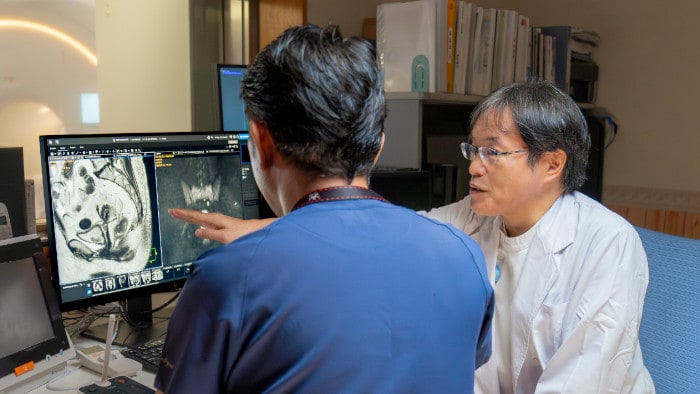
At Kumamoto Chuo Hospital, Japan, Dr. Kazuhiro Katahira has been achieving fast, high quality images with AI-enabled SmartSpeed MRI technology. In use for six months, he describes the image quality improvements that he has seen in imaging of the whole body, spine, abdomen, prostate, breast and more. He also highlights the great impact of motion-free imaging in upper abdominal and prostate exams.

In my experience, diagnosis has clearly become easier since we started using SmartSpeed. In many instances we have been able to quickly find answers to cases that we had been struggling with in the past.
Dr. Kazuhiro Katahira
Radiologist at Kumamoto Chuo Hospital, focusing on diagnostic MRI, CT and RI, with an interest in IVR.
Impressed by AI-enabled improvements
For Kumamoto Chuo Hospital, Japan, using SmartSpeed technology was their first experience with AI-enabled MRI acquisition. “Before that, we had been using Compressed SENSE, which was a first significant step forward in speed and quality compared to SENSE that we used before,” says Dr. Kazuhiro Katahira, Radiologist at Kumamoto Chuo Hospital. He notes that after the considerable image quality improvement with Compressed SENSE, adopting AI-enabled SmartSpeed has taken fast, high-quality imaging to the next level. “I like using SmartSpeed because our image quality has considerably improved and the time required for imaging has shortened, making it a very useful technology,” he says. “In my experience, diagnosis has clearly become easier since we started using SmartSpeed. In many instances we have been able to quickly find answers to cases that we had been struggling with in the past.”
At 6 months, SmartSpeed demonstrates outstanding versatility
Kumamoto Chuo Hospital performs 7000 to 8000 MRI examinations annually. These include a large number of body imaging exams, including approximately 1200 prostate exams, 700 to 800 whole body diffusion exams, as well as liver, bile and fluid exams, and in special cases imaging of the chest area. The hospital uses two Philips MRI machines, a 3.0T Elition and a 1.5T Ingenia. When SmartSpeed was first employed, it was clear that its AI aspect would be particularly useful. Dr. Katahira explains that it improves the SNR (signal-to-noise ratio) basically by denoising, to create beautiful images. He has identified many advantages. “When higher SNR is not needed, the improved quality can be traded for other benefits, such as shortening the scan time, increasing spatial resolution, sharpening the image quality and many other applications, making it clinically very useful,” he says. After six months of use, “SmartSpeed is incorporated into all of our examinations. Its clinical advantages are very difficult to sum up in a few words,” according to Dr. Katahira, so he highlights some specific examples: “It helps us in the diagnosis of breast cancer by showing great detail, allowing us to observe development in the breast duct. And in the diagnosis of rectal cancer, it helps us see the invasion of the cancer and the surrounding area in greater detail.”
In the diagnosis of rectal cancer, it helps us see the invasion of the cancer and the surrounding area in greater detail.
Dr. Kazuhiro Katahira
Radiologist at Kumamoto Chuo Hospital, Japan
Faster lumbar spine exams are beneficial for patients with pain
Many patients who must undergo a lumbar spine examination suffer from back pain. For these patients it is difficult to maintain the imaging position long enough to successfully complete the examination. “In such cases, using SmartSpeed allows us to perform volume imaging, so that we acquire only one high resolution 3D sequence in a short time and then reconstruct the other orientations from that,” Dr. Katahira says. “This is highly advantageous because the patient needs only endure a short exam time, whereas before it was necessary to acquire a larger number of sequences in total. We have seen that the shorter time has allowed us to scan patients who previously could not finish the exam. This is a great advantage.”
Fast lumbar spine imaging for successful exam of patient in pain
A patient arrived saying that undergoing MRI was not possible because of severe back pain and leg pain, was imaged with SmartSpeed in only 94 seconds. The scan was diagnostic and afterwards the patient confirmed that it only took a little while. Performed on Elition X.
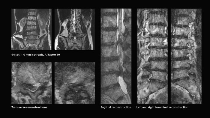
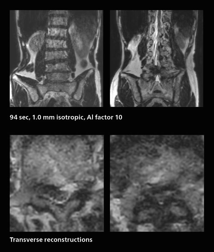
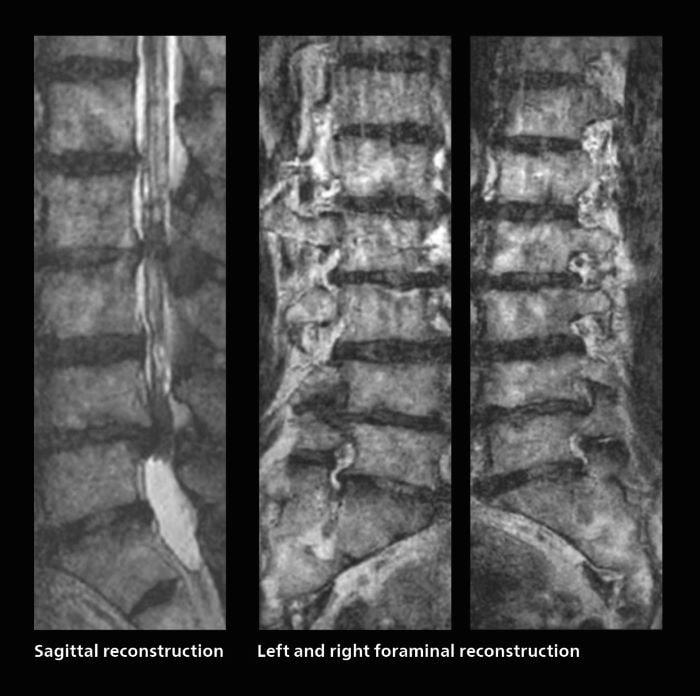
The hospital’s fast lumbar spine ExamCard includes T2W SpineVIEW, 1:40 min, 1.0 mm isotropic, acceleration factor 12.
Download the Lumbar spine SmartSpeed ExamCard from Dr. Katahira (Elition X, Release 11)
Fast, dynamic breast imaging for diagnostic confidence
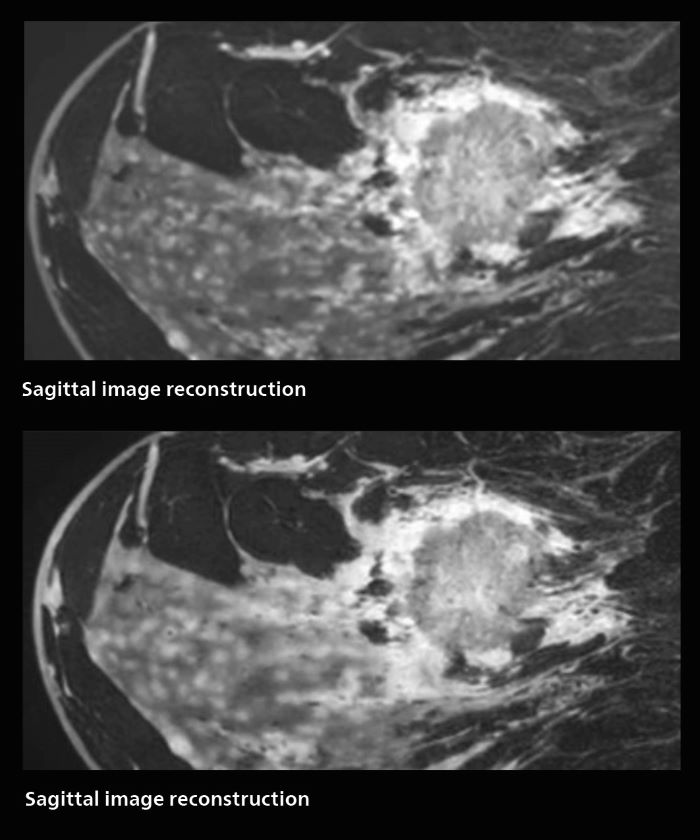
Speed and high image quality are also important factors determining the diagnostic value of breast MRI. “When the spatial resolution is not high enough for making the diagnosis of breast cancer, a very difficult decision must be made,” says Dr. Katahira. “Since SmartSpeed now allows us to increase the resolution, we can often easily provide a confident answer. In the past with SENSE we used 1.2 mm isotropic voxels in breast imaging after contrast admission. With Compressed SENSE that is 0.8 mm. Now with SmartSpeed we can acquire 0.6 mm isotropic voxels and the images are so clear that even tiny details are clearly visible.” “For example, we can now scan 20 consecutive, very fast dynamic images of the mammary glands with a single 3-second volume acquisition. This allows us to see how the blood flow is progressing in a very different way.” “The use of SmartSpeed has considerably improved our breast cancer imaging, with higher temporal resolution, higher spatial resolution, and higher SNR compared to the past, when we were using just Compressed SENSE. In addition, the dynamic study is now more useful in diagnosis because the ultrafast dynamic scan can be taken every 3 seconds.”
SmartSpeed has considerably improved our breast cancer imaging, with higher temporal resolution, higher spatial resolution, and higher SNR compared to the past.
Dr. Kazuhiro Katahira
Radiologist at Kumamoto Chuo Hospital, Japan
3D MRI of breast cancer
Scanning was performed with two different voxel sizes. AI enabled volume MRI allows image reconstruction in other directions. Biopsy revealed invasive ductal carcinoma in this patient. Performed on Elition X.
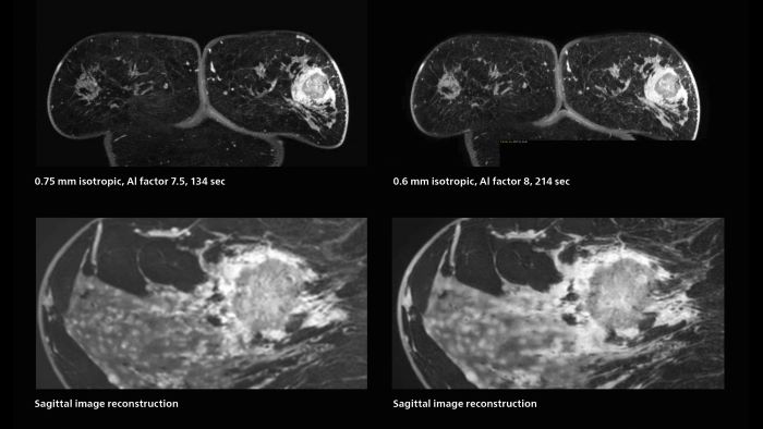
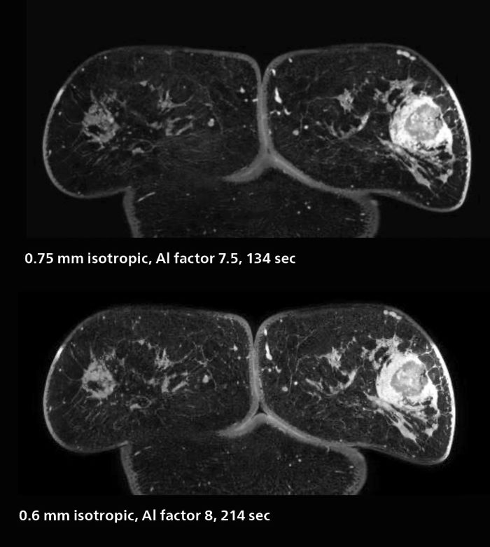
The hospital’s routine ExamCards for breast imaging include:
Download the Breast SmartSpeed ExamCard from Dr. Katahira (Elition X, Release 11)
Notable improvement in diffusion-weighted imaging
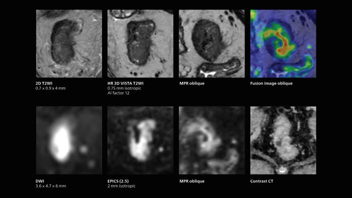
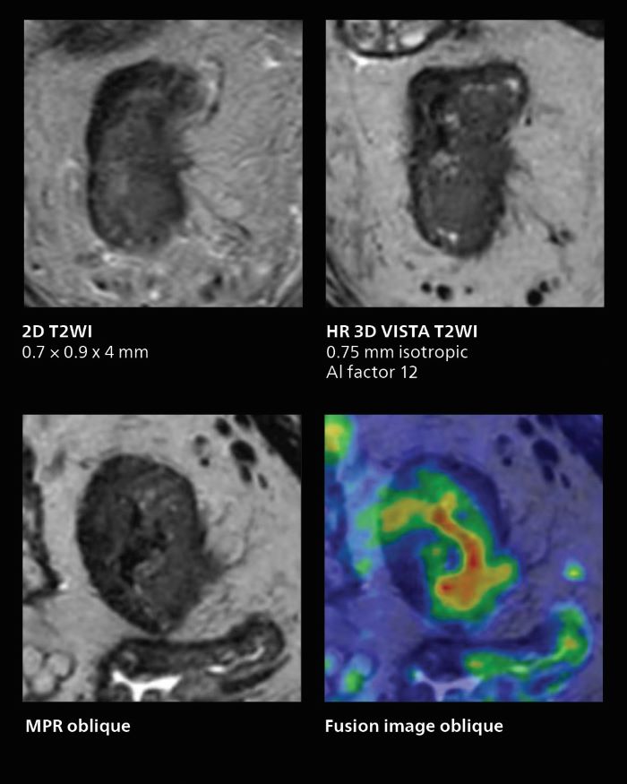
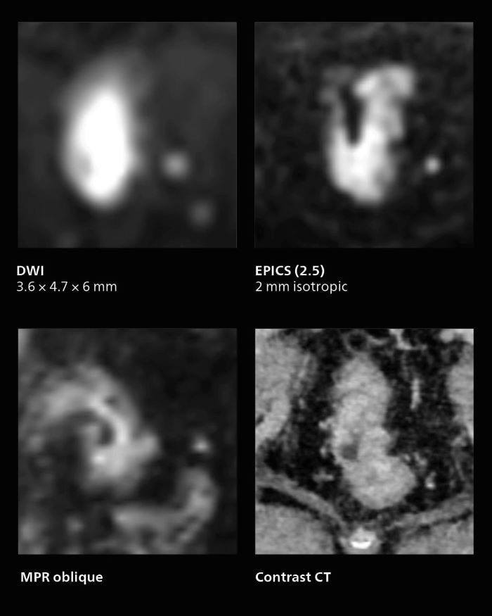
With SmartSpeed, Kumamoto Chuo Hospital also has the ability to use EPI diffusion-weighted imaging (EPICS-DWI) with Compressed SENSE, which is an important step forward according to Dr. Katahira. “Before, our EPI diffusion was performed using SENSE, but now with Compressed SENSE it is possible to obtain very clear images,” he says. He also describes the benefit of being able to perform 3D diffusion-weighted imaging. “Previously, we only had DWI images in one direction to make a diagnosis. Now, we can do something that was not possible before: performing a DWI volume acquisition so that multiplanar reconstruction can be used, allowing us to look at scan results from all directions to make the diagnosis,” Dr. Katahira says. “What used to be a diagnosis based on just cross-sectional images, can now be based on a volume image. This is a dramatic improvement for us, because it is now possible to look at slices in various cross section directions. For example, the presence or absence of venous invasion is very important in rectal cancer patients, because venous invasion can cause metastasis in the future. The ability to reconstruct images according to the direction of the blood vessels, allows us to see venous infiltration more realistically, which is a world of difference from what we were used to.”
Now, we can do something not possible before: a DWI volume acquisition so that multiplanar reconstruction allows us to look at scan results from all directions to make the diagnosis.
Dr. Kazuhiro Katahira
Radiologist at Kumamoto Chuo Hospital, Japan
MRI of rectal cancer
In this patient MRI was done to help in diagnosing the depth of invasion. Performed on Elition X.
The hospital’s routine ExamCard for rectal cancer includes:
Download the Pelvis rectal colon SmartSpeed ExamCard from Dr. Katahira (Elition X, Release 11)
Prostate imaging with SmartSpeed
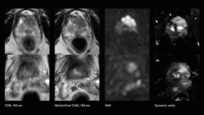
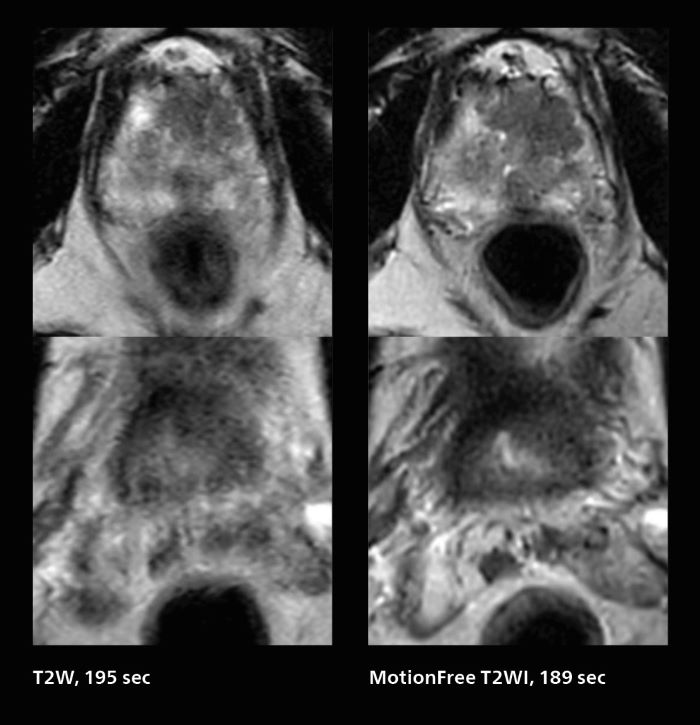
Given the high volume of prostate MRI performed at Kumamoto Chuo Hospital, the additional capabilities that SmartSpeed brings have a significant impact. “The clinical usefulness of SmartSpeed is clearly demonstrated in this area,” says Dr. Katahira. “Including SmartSpeed in most of our MRI protocols helps us improve the image quality.” The MRI team performs well over 20 prostate MRI exams per week. “In T2-weighted images we can get higher resolution,” says Dr. Katahira. “And while previously we were used to seeing blurred images due to rectal movements, introduction of the MotionFree SmartSpeed application allows us to obtain beautiful motion-compensated images, that are very useful for diagnosing, which was previously often impossible.” “In the diffusion-weighted images, we can use Compressed SENSE to obtain high quality images, and AI can be added to further improve image quality,” Dr. Katahira says.
SmartSpeed MotionFree allows us to obtain beautiful motion-compensated images, that are very useful for diagnosing, which was previously often impossible.
Dr. Kazuhiro Katahira
Radiologist at Kumamoto Chuo Hospital, Japan
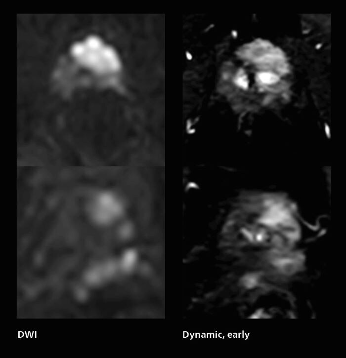
Prostate cancer MRI
MRI was performed in a patient with PSA 89.2. Evaluation of T2WI images was difficult due to rectal peristalsis. Using SmartSpeed MotionFree T2WI provided very good imaging quality. Seminal vesicle gland invasion is easily seen. Biopsy resulted in GS4+5=9. Performed on Elition X.
The hospital’s routine ExamCard for prostate cancer includes:
Download the Prostate SmartSpeed ExamCard from Dr. Katahira (Elition X, Release 11)
MotionFree abdominal scans
SmartSpeed helps achieve imaging that used to be problematic before. “When patients have difficulty holding their breath, for example in upper abdominal examinations, it is often not possible to perform an MRI examination,” says Dr Katahira. “However, with SmartSpeed we now have various ways to accomplish it. A direct benefit to these patients is the shorter scan time possible. The motion-free techniques allow us to now acquire clean single shot images. We have seen that this helps many such patients complete the examination.”
For patients having difficulty holding their breath, a direct benefit is the shorter scan time possible.
Dr. Kazuhiro Katahira
Radiologist at Kumamoto Chuo Hospital, Japan
Images of the upper abdomen can easily get blurred due to the patient’s breathing. Before SmartSpeed a viable alternative method was lacking. “We like to use single shot T2-weighted images but in the past these would look like heavy T2-weighted images, which is why we added halfscan, but this often introduced blurring,” says Dr. Katahira. “So there was a limit to what could be done. Now, with SmartSpeed, the reduction factor can be increased, so halfscan can be turned off, and sharp images can be obtained. A single-shot T2-weighted image can be taken in about 10 seconds. Because of their high resistance to movement, acquiring multiple of these images can be a substitute, even if there is some patient motion. So, thanks to SmartSpeed, we can now obtain sharp single shot images without halfscan, making single shot a good alternative to multishot.”
Rapid clarity for stroke and liver
For patients with acute cerebral infarction, who cannot endure long examinations, some clear benefits of SmartSpeed emerge. “We have a fast MR scan protocol that quickly completes the scanning. It allows us to acquire all the images we need in about 3 to 5 minutes,” says Dr. Katahira. “And it is often possible to acquire more images at that level, which is an advantage because the exam is complete even if the patient moves in the middle of the exam.” Also in dynamic MRI of the liver Dr. Katahira sees important improvements. While previously his scan used 9 seconds for a 5 mm slice, SmartSpeed now allows him to achieve a thin slice volume scan (1.6 x 2.1 x 2mm) with double arterial phase using acceleration factor 8. He indicates this is very useful for the radiologist when diagnosing, especially because it can provide a high temporal resolution.
For patients with acute cerebral infarction, we have a fast protocol that allows us to acquire all the images we need in about 3 to 5 minutes.
Dr. Kazuhiro Katahira
Radiologist at Kumamoto Chuo Hospital, Japan
Dynamic MRI of liver using SmartSpeed
A patient was referred for MR imaging of HCC. A double arterial volume dynamic study was performed. Since it is a volume dynamic study, it can also be evaluated using MPR images. Performed on Elition X.
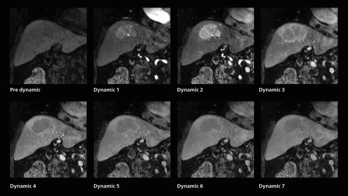
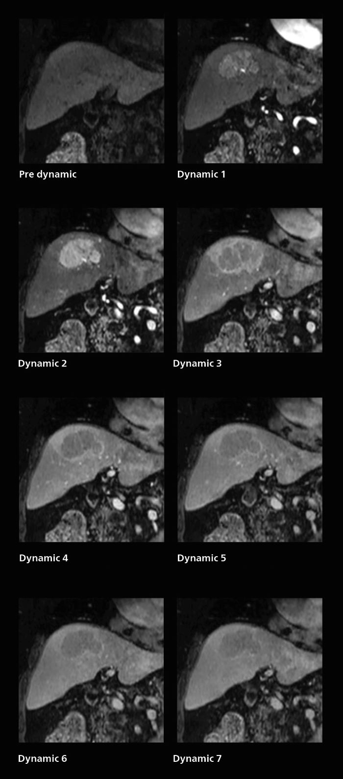
The hospital’s routine ExamCard for dynamic MRI of the liver uses total scan duration 1:05 min, dynamic scan time 9.2 sec, 1.6 x 1.8 x 2.0 mm, 200 slices, acceleration factor 8.
Download the Liver EOB Routine ExamCard from Dr. Katahira (Elition X, Release 11)
Results from case studies are not predictive of results in other cases. Results in other cases may vary.
Share this article
Register for Radiology Ingishts newsletter Our periodic Radiology Ingishts newsletter provides you articles on user experiences and best practices. Subscribe now to receive our free Radiology Ingishts newsletter via e-mail.

