SmartCT Angio

Imaging technology

Find similar products

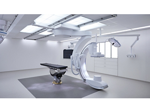
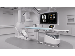
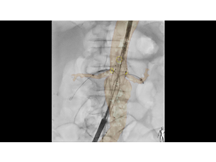
This X-ray acquisition technique generates a complete high-resolution 3D visualization of cerebral, cardiac, abdominal or peripheral vasculature from a single rotational angiography run – all controlled via the touch screen at the table. This can improve visibility of tortuous or complex anatomy that may not be seen on a 2D or DSA image.

Features
3D imaging at table side

3D imaging at table side

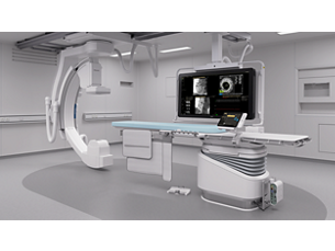
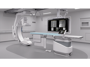
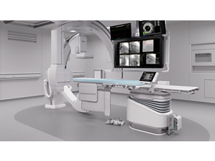
With the touch screen, you can easily acquire 3D images and interact with SmartCT tools in a natural and effortless way. Once acquired, the SmartCT viewing application automatically opens your 3D image with the correct rendering and viewing tools on the touch screen module. All tools work with touch screen simplicity within the sterile area.

3D imaging at table side

3D imaging at table side
With the touch screen, you can easily acquire 3D images and interact with SmartCT tools in a natural and effortless way. Once acquired, the SmartCT viewing application automatically opens your 3D image with the correct rendering and viewing tools on the touch screen module. All tools work with touch screen simplicity within the sterile area.

3D imaging at table side

With the touch screen, you can easily acquire 3D images and interact with SmartCT tools in a natural and effortless way. Once acquired, the SmartCT viewing application automatically opens your 3D image with the correct rendering and viewing tools on the touch screen module. All tools work with touch screen simplicity within the sterile area.
Click here for more information
3D imaging at table side

3D imaging at table side

With the touch screen, you can easily acquire 3D images and interact with SmartCT tools in a natural and effortless way. Once acquired, the SmartCT viewing application automatically opens your 3D image with the correct rendering and viewing tools on the touch screen module. All tools work with touch screen simplicity within the sterile area.
Reveal information not apparent on 2D or DSA images

Reveal information not apparent on 2D or DSA images

SmartCT offers advanced reconstruction techniques to improve anatomical visualization. Once acquired, you can manipulate advanced visualizations such as 3D MPR on the table side touch screen to evaluate the type and extent of disease and surrounding tissues with great detail. SmartCT 3D images can help reveal information not apparent on 2D or DSA images, which can impact clinical decisions.

Reveal information not apparent on 2D or DSA images

Reveal information not apparent on 2D or DSA images
SmartCT offers advanced reconstruction techniques to improve anatomical visualization. Once acquired, you can manipulate advanced visualizations such as 3D MPR on the table side touch screen to evaluate the type and extent of disease and surrounding tissues with great detail. SmartCT 3D images can help reveal information not apparent on 2D or DSA images, which can impact clinical decisions.

Reveal information not apparent on 2D or DSA images

SmartCT offers advanced reconstruction techniques to improve anatomical visualization. Once acquired, you can manipulate advanced visualizations such as 3D MPR on the table side touch screen to evaluate the type and extent of disease and surrounding tissues with great detail. SmartCT 3D images can help reveal information not apparent on 2D or DSA images, which can impact clinical decisions.
Click here for more information
Reveal information not apparent on 2D or DSA images

Reveal information not apparent on 2D or DSA images

SmartCT offers advanced reconstruction techniques to improve anatomical visualization. Once acquired, you can manipulate advanced visualizations such as 3D MPR on the table side touch screen to evaluate the type and extent of disease and surrounding tissues with great detail. SmartCT 3D images can help reveal information not apparent on 2D or DSA images, which can impact clinical decisions.
SmartCT Vessel Analysis supports treatment planning

SmartCT Vessel Analysis supports treatment planning

Select two points of a vessel to quickly define a vessel path on a 3D volume. The path is automatically detected and rendered in different views to support easy inspection of vessel and device positioning with straightened, curved and cross-section reformats. The vessel dimensions are automatically extracted, and landing zones can be specified and later overlaid on live fluoroscopy. Finally, optimal projection angles for catheterization can be identified and later recalled with a single button press.

SmartCT Vessel Analysis supports treatment planning

SmartCT Vessel Analysis supports treatment planning
Select two points of a vessel to quickly define a vessel path on a 3D volume. The path is automatically detected and rendered in different views to support easy inspection of vessel and device positioning with straightened, curved and cross-section reformats. The vessel dimensions are automatically extracted, and landing zones can be specified and later overlaid on live fluoroscopy. Finally, optimal projection angles for catheterization can be identified and later recalled with a single button press.

SmartCT Vessel Analysis supports treatment planning

Select two points of a vessel to quickly define a vessel path on a 3D volume. The path is automatically detected and rendered in different views to support easy inspection of vessel and device positioning with straightened, curved and cross-section reformats. The vessel dimensions are automatically extracted, and landing zones can be specified and later overlaid on live fluoroscopy. Finally, optimal projection angles for catheterization can be identified and later recalled with a single button press.
Click here for more information
SmartCT Vessel Analysis supports treatment planning

SmartCT Vessel Analysis supports treatment planning

Select two points of a vessel to quickly define a vessel path on a 3D volume. The path is automatically detected and rendered in different views to support easy inspection of vessel and device positioning with straightened, curved and cross-section reformats. The vessel dimensions are automatically extracted, and landing zones can be specified and later overlaid on live fluoroscopy. Finally, optimal projection angles for catheterization can be identified and later recalled with a single button press.
Easily perform two-point measurements on screen

Easily perform two-point measurements on screen

Quickly measure distances on a 2D or 3D image with two-point measurements on the touch screen. This can help quickly check the trajectory to a target vessel, measure distances for stent deployment, measure the size of anatomy or identify a discrepancy to speed up planning of the optimal treatment angle and aid navigation.

Easily perform two-point measurements on screen

Easily perform two-point measurements on screen
Quickly measure distances on a 2D or 3D image with two-point measurements on the touch screen. This can help quickly check the trajectory to a target vessel, measure distances for stent deployment, measure the size of anatomy or identify a discrepancy to speed up planning of the optimal treatment angle and aid navigation.

Easily perform two-point measurements on screen

Quickly measure distances on a 2D or 3D image with two-point measurements on the touch screen. This can help quickly check the trajectory to a target vessel, measure distances for stent deployment, measure the size of anatomy or identify a discrepancy to speed up planning of the optimal treatment angle and aid navigation.
Click here for more information
Easily perform two-point measurements on screen

Easily perform two-point measurements on screen

Quickly measure distances on a 2D or 3D image with two-point measurements on the touch screen. This can help quickly check the trajectory to a target vessel, measure distances for stent deployment, measure the size of anatomy or identify a discrepancy to speed up planning of the optimal treatment angle and aid navigation.
Quickly define any structure of interest

Quickly define any structure of interest

The SmartCT semi-automatic lesion segmentation tool allows you to easily define any structure of interest, measure its volume and remove anatomy to improve visualization.

Quickly define any structure of interest

Quickly define any structure of interest
The SmartCT semi-automatic lesion segmentation tool allows you to easily define any structure of interest, measure its volume and remove anatomy to improve visualization.

Quickly define any structure of interest

The SmartCT semi-automatic lesion segmentation tool allows you to easily define any structure of interest, measure its volume and remove anatomy to improve visualization.
Click here for more information
Quickly define any structure of interest

Quickly define any structure of interest

The SmartCT semi-automatic lesion segmentation tool allows you to easily define any structure of interest, measure its volume and remove anatomy to improve visualization.
  • 3D imaging at table side
  • Reveal information not apparent on 2D or DSA images
  • SmartCT Vessel Analysis supports treatment planning
  • Easily perform two-point measurements on screen
See all features
3D imaging at table side

3D imaging at table side

With the touch screen, you can easily acquire 3D images and interact with SmartCT tools in a natural and effortless way. Once acquired, the SmartCT viewing application automatically opens your 3D image with the correct rendering and viewing tools on the touch screen module. All tools work with touch screen simplicity within the sterile area.

3D imaging at table side

3D imaging at table side
With the touch screen, you can easily acquire 3D images and interact with SmartCT tools in a natural and effortless way. Once acquired, the SmartCT viewing application automatically opens your 3D image with the correct rendering and viewing tools on the touch screen module. All tools work with touch screen simplicity within the sterile area.

3D imaging at table side

With the touch screen, you can easily acquire 3D images and interact with SmartCT tools in a natural and effortless way. Once acquired, the SmartCT viewing application automatically opens your 3D image with the correct rendering and viewing tools on the touch screen module. All tools work with touch screen simplicity within the sterile area.
Click here for more information
3D imaging at table side

3D imaging at table side

With the touch screen, you can easily acquire 3D images and interact with SmartCT tools in a natural and effortless way. Once acquired, the SmartCT viewing application automatically opens your 3D image with the correct rendering and viewing tools on the touch screen module. All tools work with touch screen simplicity within the sterile area.
Reveal information not apparent on 2D or DSA images

Reveal information not apparent on 2D or DSA images

SmartCT offers advanced reconstruction techniques to improve anatomical visualization. Once acquired, you can manipulate advanced visualizations such as 3D MPR on the table side touch screen to evaluate the type and extent of disease and surrounding tissues with great detail. SmartCT 3D images can help reveal information not apparent on 2D or DSA images, which can impact clinical decisions.

Reveal information not apparent on 2D or DSA images

Reveal information not apparent on 2D or DSA images
SmartCT offers advanced reconstruction techniques to improve anatomical visualization. Once acquired, you can manipulate advanced visualizations such as 3D MPR on the table side touch screen to evaluate the type and extent of disease and surrounding tissues with great detail. SmartCT 3D images can help reveal information not apparent on 2D or DSA images, which can impact clinical decisions.

Reveal information not apparent on 2D or DSA images

SmartCT offers advanced reconstruction techniques to improve anatomical visualization. Once acquired, you can manipulate advanced visualizations such as 3D MPR on the table side touch screen to evaluate the type and extent of disease and surrounding tissues with great detail. SmartCT 3D images can help reveal information not apparent on 2D or DSA images, which can impact clinical decisions.
Click here for more information
Reveal information not apparent on 2D or DSA images

Reveal information not apparent on 2D or DSA images

SmartCT offers advanced reconstruction techniques to improve anatomical visualization. Once acquired, you can manipulate advanced visualizations such as 3D MPR on the table side touch screen to evaluate the type and extent of disease and surrounding tissues with great detail. SmartCT 3D images can help reveal information not apparent on 2D or DSA images, which can impact clinical decisions.
SmartCT Vessel Analysis supports treatment planning

SmartCT Vessel Analysis supports treatment planning

Select two points of a vessel to quickly define a vessel path on a 3D volume. The path is automatically detected and rendered in different views to support easy inspection of vessel and device positioning with straightened, curved and cross-section reformats. The vessel dimensions are automatically extracted, and landing zones can be specified and later overlaid on live fluoroscopy. Finally, optimal projection angles for catheterization can be identified and later recalled with a single button press.

SmartCT Vessel Analysis supports treatment planning

SmartCT Vessel Analysis supports treatment planning
Select two points of a vessel to quickly define a vessel path on a 3D volume. The path is automatically detected and rendered in different views to support easy inspection of vessel and device positioning with straightened, curved and cross-section reformats. The vessel dimensions are automatically extracted, and landing zones can be specified and later overlaid on live fluoroscopy. Finally, optimal projection angles for catheterization can be identified and later recalled with a single button press.

SmartCT Vessel Analysis supports treatment planning

Select two points of a vessel to quickly define a vessel path on a 3D volume. The path is automatically detected and rendered in different views to support easy inspection of vessel and device positioning with straightened, curved and cross-section reformats. The vessel dimensions are automatically extracted, and landing zones can be specified and later overlaid on live fluoroscopy. Finally, optimal projection angles for catheterization can be identified and later recalled with a single button press.
Click here for more information
SmartCT Vessel Analysis supports treatment planning

SmartCT Vessel Analysis supports treatment planning

Select two points of a vessel to quickly define a vessel path on a 3D volume. The path is automatically detected and rendered in different views to support easy inspection of vessel and device positioning with straightened, curved and cross-section reformats. The vessel dimensions are automatically extracted, and landing zones can be specified and later overlaid on live fluoroscopy. Finally, optimal projection angles for catheterization can be identified and later recalled with a single button press.
Easily perform two-point measurements on screen

Easily perform two-point measurements on screen

Quickly measure distances on a 2D or 3D image with two-point measurements on the touch screen. This can help quickly check the trajectory to a target vessel, measure distances for stent deployment, measure the size of anatomy or identify a discrepancy to speed up planning of the optimal treatment angle and aid navigation.

Easily perform two-point measurements on screen

Easily perform two-point measurements on screen
Quickly measure distances on a 2D or 3D image with two-point measurements on the touch screen. This can help quickly check the trajectory to a target vessel, measure distances for stent deployment, measure the size of anatomy or identify a discrepancy to speed up planning of the optimal treatment angle and aid navigation.

Easily perform two-point measurements on screen

Quickly measure distances on a 2D or 3D image with two-point measurements on the touch screen. This can help quickly check the trajectory to a target vessel, measure distances for stent deployment, measure the size of anatomy or identify a discrepancy to speed up planning of the optimal treatment angle and aid navigation.
Click here for more information
Easily perform two-point measurements on screen

Easily perform two-point measurements on screen

Quickly measure distances on a 2D or 3D image with two-point measurements on the touch screen. This can help quickly check the trajectory to a target vessel, measure distances for stent deployment, measure the size of anatomy or identify a discrepancy to speed up planning of the optimal treatment angle and aid navigation.
Quickly define any structure of interest

Quickly define any structure of interest

The SmartCT semi-automatic lesion segmentation tool allows you to easily define any structure of interest, measure its volume and remove anatomy to improve visualization.

Quickly define any structure of interest

Quickly define any structure of interest
The SmartCT semi-automatic lesion segmentation tool allows you to easily define any structure of interest, measure its volume and remove anatomy to improve visualization.

Quickly define any structure of interest

The SmartCT semi-automatic lesion segmentation tool allows you to easily define any structure of interest, measure its volume and remove anatomy to improve visualization.
Click here for more information
Quickly define any structure of interest

Quickly define any structure of interest

The SmartCT semi-automatic lesion segmentation tool allows you to easily define any structure of interest, measure its volume and remove anatomy to improve visualization.

You are about to visit a Philips global content page

Continue

You are about to visit a Philips global content page

Continue

Our site can best be viewed with the latest version of Microsoft Edge, Google Chrome or Firefox.

Jūs pateksite į Lietuvos „Philips Healthcare“ svetainę.
Please select the checkbox

Svetainė skirta tik asmenims, naudojantiems medicinos priemones profesinėje veikloje, įskaitant sveikatos priežiūros specialistams, profesinę veiklą vykdantiems asmenims, atstovaujantiems sveikatos priežiūros įstaigą ar medicinos priemonėmis prekiaujančią organizaciją.

Ar esate asmuo, atitinkantis aukščiau nurodytus kriterijus?