
No bounds. Better healthcare.
Measuring pediatric and adult tumors using 3D imaging
3D imaging technology advances radiology clinical processes
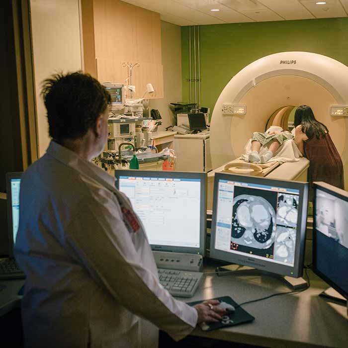
Diagnostic imaging has come a long way from low-resolution images of the human anatomy and days-long post-processing. In 1999, it could take five days1 for computed tomographic (CT) images to be turned around. Today, a radiologist can scan a baby’s vital organs and see a 3D reconstruction within seconds. With 3D printing capabilities, those images can then be used to provide a 360-degree view of a patient’s heart. In the past three decades pediatric radiology has been transformed by imaging technology. More simplicity, speed and accuracy has allowed improved workflows, more accurate measurements and faster clinical decisions, but likely nothing has opened up as many new opportunities as the transition from 2D to 3D imaging. Phoenix Children’s Hospital has been using 3D imaging for several years, and has incorporated it into several areas of its patient care, such as preoperative planning, research and education, and patient and family support, including its pediatric patients. “We’ve made a big effort in the department of radiology to better present imaging data to the users of that information,” says Dr. Richard Towbin, Chief of Radiology at Phoenix Children’s. “Any time we get digital data we can morph it into 3D, display it in 3D, and then, with the materials and unique data that we’re getting, make decisions which are going to improve the clinical outcome.”
The conversion from 2D to 3D
The journey to a virtual 3D image which is displayed on a computer screen begins with a CT or MRI scan, from which the images are processed through Philips IntelliSpace Portal advanced visualization software, allowing radiologists to take 2D and 3D images, segment images, and efficiently take measurements and diagnose more rapidly than before. “Getting to that 3D print takes a lot of technology. That technology begins with the Philips CT or MR scan, and proceeds through the IntelliSpace Portal, where there is some very technical image segmentation that goes on in our 3D Lab. It takes a little bit of time but really the technology is there and if I couldn’t see it and rely upon the segmentation technology of the IntelliSpace Portal, then we would not be able to get anywhere,” says Dr. Dianna Bardo, Director of Body MR & Co-Director of 3D Innovation Lab at Phoenix Children’s. Tumors, for example, are notoriously difficult to measure given their unusual sizes and shapes. Current protocols call for measuring tumors using length, width and height data. “First of all, a clinician may not be able to look at a 2-dimensional image and get a real, accurate idea of what a disease process looks like, whether that’s in the heart, in the liver, or in a fractured bone,” says Dr. Bardo. “We’ve had 11 radiologists measure 100 tumors in 100 different patients, and everybody’s perception of the shape of the tumor varies considerably. The linear measurements they make vary considerably, sometimes as much as a liter difference in the volume of a tumor, all measured from the same imaging.”
Applications for medical 3D printing in radiology
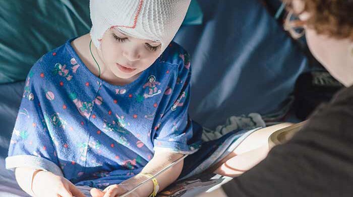
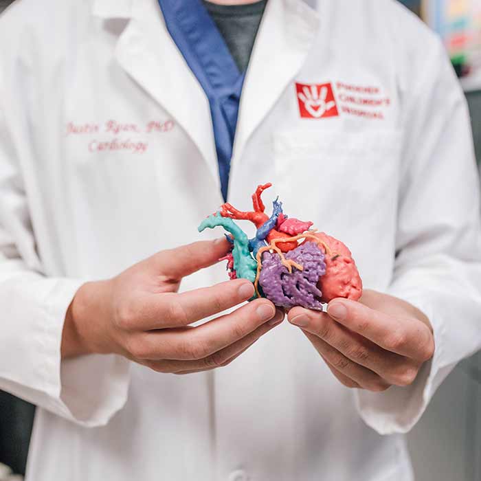
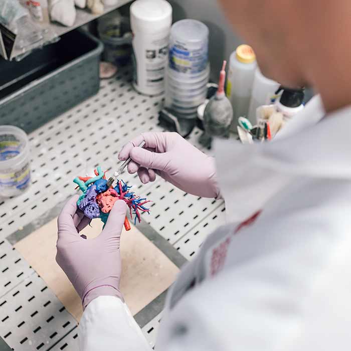
Using 3D technology, radiologists at Phoenix Children’s can determine a tumor’s exact size and physicians can discern subtle changes in size or shape over time and treatment. The precision of 3D imaging allows oncologists to identify the most viable course of treatment and determine whether treatment is shrinking a patient’s tumor. “When I model a tumor or model a heart or bone into a 3-dimensional object in our 3D Lab, it’s an instant picture. It’s something anybody can perceive,” says Dr. Bardo. “A patient can perceive it. Their parents can perceive it well and understand the disease and proposed treatment more readily. The physician that’s caring for them, the surgeon that may be repairing something, and the radiologists all have this understanding.” Innovation in image processing and the evolution from 2D to 3D has led to greater accuracy. The 3D Innovation Lab at Phoenix Children’s develops images that are translated by the Hospital’s Cardiac 3D Print Lab to incorporate advanced post-processing technologies. These models help prepare physicians for complex procedures by creating accurate models of defective hearts, limb deficiencies, injured internal organs, and even tumors. Cardiovascular surgeons, for example, can view individual segment parts of the heart using a 3D image, or can hold a 3D print of the heart in their hands. Both can help them gain a better understanding of complex anatomy prior to surgery. “We process the images in radiology, reconstruct and segment the anatomy, and then send it electronically to our Cardiac 3D Print Lab. If it’s necessary for the surgeon or patient-family to better understand the anatomy, we can make a 3D printed image of the heart. We can allow a doctor or a family to hold a heart in the palm of their hand,” Justin Ryan, Research Scientist of the Cardiac 3D Print Lab at Phoenix Children’s. The models also help families understand their infant’s surgery and help train medical students and residents. “We have so many uses for these 3D printed models. The child can take it to school for show-and-tell, bring it to the OR for planning, or carry it to the clinic for family education. It’s all very integrated into the child’s life from a holistic care perspective, and it all starts in radiology,” says Dr. Ryan.
Improving hospital efficiency and reducing waste
With hospitals looking to reduce waste in their operations, diagnostic imaging is one area of potential efficiency. With CT scans in the US nearly tripling from 52 scans per 1,000 patients to 149 scans per 1,000 patients between 1996 and 2010, Dr. Towbin says the transition from 2D to 3D has shown the potential to reduce the number of repeat scans at Phoenix Children’s Hospital because care providers are able to glean more information and understand it more readily from every scan. “All the technology, information technology, business technology and analysis that Philips helps us provide allows us to better look at our operations and procedures and find areas that we’re inefficient at doing, and when we find those things, then we have an opportunity to modify the process and thereby reduce our waste or inefficiency,” Dr. Towbin says. “3D is a great example of that because now we have to do less imaging because we’ve answered questions that previously we weren’t able to answer with the 2D data.”
See how Phoenix Children's Hospital is
Erasing boundaries in pediatric radiology
Getting to that 3D print takes a lot of technology. That technology begins with the Philips CT or MR scan, and proceeds through the IntelliSpace Portal.”
Chief of Radiology at Phoenix
Dr. Richard Towbin
Children's Hospital
Advanced visualization with Philips IntelliSpace Portal
No bounds.
Better healthcare.
There's always a way to make life better.
DISCLAIMER: Results are specific to the institution where they were obtained and may not reflect the results achievable at other institutions. FOOTNOTES:
1 Radiographics, ‘Radiologic Professionalism in Modern Health Care’, Anastasia L. Hryhorczuk, MD, Kate Hanneman, MD1, Ronald L. Eisenberg, MD, JD, Elaine C. Meyer, RN, PhD, Stephen D. Brown, MD